Jun 21, 2016Think of a digital image that is the size of a dining room table. Now consider scanning that image for a particular color or pattern. There is a great deal of space to survey. But the individual who needs to analyze that image need not be present in the dining room.
That's how Deepak Puri, the chief solutions architect for a San Francisco-based technology consultancy called Skilled Analysts, explains digital pathology. What undergirds this process is virtual microscopy, technology that allows a pathologist to remotely view a blood or tissue specimen on a glass slide on a microscope that creates a high-resolution image, which is then saved in the cloud.
The digital microscopes needed for this process are extremely costly and their use requires reliable access to high-bandwidth Internet, in order for the full image (the entire dining room table, as noted above) to be accessible. At the other end of the technology spectrum are cheap zoom lenses that can be attached to a smartphone to capture an image of the slide. But the detail in the imagery may be inadequate for a pathologist at a remote location to make a diagnosis.
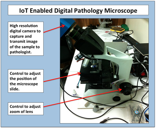
So Puri and his team have been developing a third option: a retrofit. Starting with a conventional microscope used by medical lab technicians, they attach motors, controllable via the Internet, to the knob used to adjust the lens (to zoom in and out over the image), as well as to the handle used to position the slide. The team also mounts a high-resolution digital camera over the viewer and makes it controllable via the Internet as well. By connecting these three microscope parts to a cloud-based platform via a cellular network (assuming such a network is available at the remote location), a pathologist in some other part of the world can control the microscope, take digital images of the sample and assign a diagnosis.
The most difficult aspect, Puri says, has been making the Web-based controls precise enough to enable the fine-tuning that a pathologist requires. "Moving the lens in the microscope is delicate operation," he states.
Initially, the team developed the prototype retrofitted microscope for use in Indian villages, so that clinics could call pathologists in distant cities and have them examine specimens and provide diagnoses remotely. Now, however, Puri and his team are also reaching out to aid agencies and clinics in parts of the world where the Zika virus is emerging.
If the team can partner with aid organizations and identity funding, they will be able to speed up the prototype's production and ship retrofitted microscopes to parts of the world where public health workers are scrambling to improve their capabilities to diagnose the Zika virus quickly. In those remote areas, Zika diagnosis is currently possible only by either bringing pathologists to the specimens, or by shipping those specimens to the pathologists—a process that can add a delay of several days before a diagnosis can be reached. What's more, the specimens could be lost or damaged during transit.
Puri says he and his colleagues are working on a browser-based simulation that "will allow users to try out the microscope—which is set up at a lab in India—with a blood sample on a slide. The user will be able to control the microscope functions via a Web browser."
